3D Medical Segmentation of Volumes
3D medical segmentation labels whole CT and MRI volumes at once so vessels and tumors stay connected across slices.
Why Does This Exist?
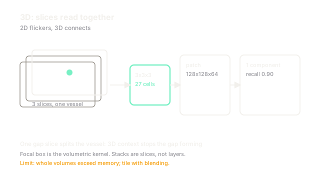
Slice models flicker: tumor present on slice , gone on , back on . Vessels disconnect. 3D nets read patches with true spacing, so through-plane context connects anatomy. DICOM handling must orient volumes first; nnU-Net configures the patches.
Think of It Like This
Reading a novel versus random pages
Judging a plot from random pages misses arcs that span chapters. Reading whole chapters keeps characters connected. Skimming pages is faster but loses threads.
Slices are pages and patches are chapters. The analogy stops at memory: chapters need GPUs while pages fit anywhere.
How It Actually Works
Volumes resample to median spacing, often mm isotropic for brains and mm for abdomens. Patches near with batch train Dice plus cross-entropy. Inference slides with percent overlap and Gaussian blending. Anisotropic kernels respect thick slices.
Worked example
A cm liver tumor spans slices at mm. Slice models score Dice with two gaps. A 3D patch sees all at once and reaches with zero gaps. Memory is bytes near MB per feature map times maps, which fits GB at batch .
Watch Out For
Thick slices stretched as isotropic cubes
Resampling mm slices to mm cubes invents four slices of blur that look like edema. The symptom is puffy masks on old scanners. Keep anisotropic spacing or acquire thin slices for 3D.
The Quick Version
- 3D models connect anatomy across slices that 2D flickers on.
- Spacing-aware patches near balance context and memory.
- Anisotropic kernels respect thick-slice reality.
- Do not fake isotropic cubes from mm slices; blur masquerades as disease.
- Sliding overlap plus blending removes patch seams.