BraTS Brain Tumor Scans
Multi-sequence brain MRI with voxel tumor labels, the ground for 3D medical segmentation under careful handling.
Why Does This Exist?
One MRI sequence hides tumor borders that another reveals. Brain tumor work needs aligned multi-sequence volumes plus voxel truth for whole, core and enhancing parts. BraTS supplies that challenge ground so medical 3D segmentation can compare identical region rules.
This page covers multi-sequence I/O and region scoring. Overlap math links to IoU and Dice once published, with U-Net as architecture context.
Think of It Like This
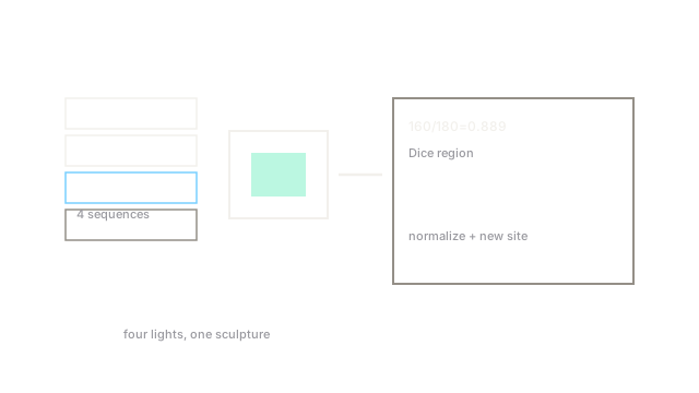
Four flashlights on the same sculpture
One flashlight hides cracks, another shows them. Four lights together reveal the full shape.
BraTS sequences are those lights. Each MRI weighting is one flashlight on the same brain.
How It Actually Works
Purpose is 3D tumor segmentation by region. Inputs are aligned MRI volumes, outputs are voxel labels for whole, core and enhancing tumor. Appropriate use is challenge research with site-generalization checks. Small structures and site shifts dominate errors, so report per region.
Toy Dice walkthrough
Take 100 truth voxels with 80 shared, 10 missed and 10 false. Dice is . Enhancing cores run smaller, so the same miss count hurts more there.
Code
Dice for one region on toy voxels.
import numpy as np
def dice(a: np.ndarray, b: np.ndarray) -> float: inter = np.sum((a == 1) & (b == 1)) return round(float(2*inter / max(a.sum() + b.sum(), 1)), 3)
print(dice(np.array([1]*80 + [0]*20), np.array([1]*80 + [1]*10 + [0]*10)))# -> 0.889Eighty shared with ten each way gives about 0.889.
Watch Out For
Tuning to one site then missing new scanners
Sequence intensity shifts by scanner, so single-site tuning drops elsewhere. The symptom is challenge strength with hospital weakness. Fix it with intensity normalization plus held-out site tests.
The Quick Version
- BraTS maps aligned brain MRI volumes to voxel tumor region labels.
- Inputs are multi-sequence scans, outputs are whole, core and enhancing masks.
- Toy 80 shared with 10 each way gives Dice about 0.889.
- Use it for 3D medical segmentation research with care.
- Normalize intensities and test held-out sites.